PHENOTHIAZINE
| Method number: | PV2048 |
| Matrix: | Air |
| Target concentration: | 5 mg/m3 (OSHA TWA PEL) |
| Procedure: | Samples are collected by drawing a known volume of air through a
glass fiber filter. Samples are extracted with methyl
|
| Air volume and sampling rate studied: |
100 liters at 1.0 liters per minute. |
| Status of method: | Stopgap method. This method has been only partially evaluated and is presented for information and trial use. |
| Date: October, 1989 | Chemist: Mary E. Eide |
SOLVENTS BRANCH
OSHA ANALYTICAL LABORATORY
SALT LAKE
CITY, UTAH
1. General Discussion
- 1.1. Background
- 1.1.1. History of procedure
The PEL for phenothiazine is 5 mg/m3. Since phenothiazine is a solid at room temperature, collection on a glass fiber filter was tried and found successful. There was no loss of phenothiazine in the retention studies. The extraction, and storage studies were near 100%.
1.1.2. Potential workplace exposure (Ref. 5.1.)
Phenothiazine is used as a pesticide, and is used orally to treat pinworm, threadworm, and roundworm infestations. It is used as a base for the manufacture of pharmaceuticals. It is used as a urinary antiseptic.
1.1.3. Toxic Effects (This section is for information purposes and should not be taken as the basis for OSHA policy.)(Ref. 5.1.)
Oral doses of 1 or more grams per day may cause toxic hepatitis,
hemolytic anemia, abdominal cramps, tachycardia, gastrointestinal
and skin irritation, skin photosensitization, kidney damage, and
pruritus. Workers applying phenothiazine in orchards reported skin
irritation, including itching, and redness on any exposed surface.
The photosensitizing dose is less than 0.75 grams. Workers exposed
to between 15 and 48 mg/m3 during
pulverizing and packaging phenothiazine dust developed
1.1.4. Physical properties (Ref. 5.2.):
| Compound: | 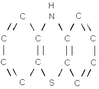 |
| Synonyms: | dibenzothiazine; Agrazine; thiodiphenylamine;
Biverm; Antiverm; Contaverm; Ieeno; ENT 38; Fenoverm;
Fentiazine; Helmetina; Lethelmin; Nemazene; Orimon; Padophene;
Penthazine; Vermitin; |
| Molecular weight: | 199.26 |
| Melting point: | 185°C |
| Boiling point: | 371°C |
| Color: | yellow rhombic leaflets or
|
| Molecular formula: | C12H9NS |
| CAS: | 92-84-2 |
| IMIS: | 2041 |
| RTECS: | 57228 (SN5075000) |
1.2. Limit defining parameters
- 1.2.1. The detection limit of the analytical procedure is 1 ng.
This is the smallest amount that could be detected under normal
operating conditions.
1.2.2. The overall detection limit is 0.03 mg/m3, based on a 3 mL extraction and a 100 liter air volume. (All mg/m3 amounts in this study are based on a 100 liter air volume and a 3 mL desorption.)
1.3. Advantages
- 1.3.1. The sampling procedure is convenient.
1.3.2. The analytical method is reproducible and sensitive.
1.3.3. Reanalysis of samples is possible.
1.3.4. It may be possible to analyze other compounds at the same time.
1.3.5. Interferences may be avoided by proper selection of column and GC parameters.
1.4. Disadvantages
none known
2. Sampling procedure
- 2.1. Apparatus
- 2.1.1. A calibrated personal sampling pump, the flow of which
can be determined within + 5% at the recommended flow.
2.1.2. A
2.2. Sampling technique
- 2.2.1. The ends of the filter cassette are opened immediately
before sampling.
2.2.2. Connect the filter cassette to the sampling pump with flexible tubing.
2.2.3. Air being sampled should not pass through any hose or tubing before entering the cassette.
2.2.4. Seal the ends of the cassette with plastic caps
immediately after sampling. Seal each sample lengthwise with OSHA
2.2.5. With each batch of samples, submit at least one blank filter from the same lot used for samples. This filter should be subjected to exactly the same handling as the samples except that no air is drawn through it.
2.2.6. Transport the samples (and corresponding paperwork) to the lab for analysis.
2.2.7. Bulks submitted for analysis must be shipped in a separate mailing container from the samples.
2.3. Extraction efficiency
Six glass fiber filters were liquid spiked at each loading of 16.56
µg (0.4968 mg/m3), 82.8 µg (2.484
mg/m3), and 165.6 µg (4.968
mg/m3) phenothiazine. They were allowed to
equilibrate overnight at room temperature. They were opened, placed
into a 4 mL vial, extracted with 3 mL of methyl
Desorption Efficiency
|
| |||
| Tube# | % Recovered | ||
| 16.56 µg | 82.8 µg | 165.6 µg | |
|
| |||
| 1 | 99.1 | 99.9 | 98.7 |
| 2 | 97.1 | 101 | 101 |
| 3 | 103 | 99.6 | lost |
| 4 | 101 | 102 | 101 |
| 5 | 99.0 | 99.4 | 101 |
| 6 | 97.9 | 98.9 | 99.6 |
| average | 100 | 100 | 99.5 |
| overall average | 99.8 | ||
| standard deviation | ± 1.51 | ||
|
| |||
2.4. Retention efficiency
Six glass fiber filters were liquid spiked with 165.6 µg (4.968
mg/m3) phenothiazine. They were placed in a
cassette with a second glass fiber filter, and a spacer between the
two filters. They were allowed to equilibrate overnight , and had 100
liters humid air (90% RH) pulled through them. They were opened,
extracted, and analyzed by
Retention Efficiency
|
| |||
| Sample # | % Recovered | % Recovered | Total |
| 'A' | 'B' | ||
|
| |||
| 1 | 99.3 | 0.0 | 99.3 |
| 2 | 97.8 | 0.0 | 97.8 |
| 3 | 101 | 0.0 | 101 |
| 4 | 98.6 | 0.0 | 98.6 |
| 5 | 100 | 0.0 | 100 |
| 6 | 98.0 | 0.0 | 98.0 |
| average | 99.1 | ||
|
| |||
2.5. Storage
Glass fiber filters were spiked with 165.6 µg (4.968 mg/m3) phenothiazine and stored at room temperature on the benchtop until opened and analyzed. Half of the storage samples were stored in brown vials, as phenothiazine decomposes in sunlight. The storage samples were exposed to room light. There was little difference between the samples stored in brown and clear glass. The spectrum of room light does not compare to sunlight, so this comparison probably does not mimic sunlight conditions. The recoveries averaged 98.9% for brown glass vials, and 99.6 % for clear glass vials for the 14 days stored (Table 3).
Storage Study
|
| ||
| Days | % Recovered | |
| brown glass | clear glass | |
|
| ||
| 6 | 102 | 102 |
| 6 | 99.7 | 99.0 |
| 6 | 100 | 101 |
| 14 | 98.3 | 98.1 |
| 14 | 97.2 | 98.2 |
| 14 | 96.2 | 99.5 |
| average | 98.9 | 99.6 |
| overall average | 99.3 | |
|
| ||
2.6. Precision
The precision was calculated using the area counts from six injections of each standard at concentrations of 16.56, 82.8, 165.6, and 331.2 µg/mL. The pooled coefficient of variation was 0.0152 (Table 4).
Precision Study
|
| ||||
| Injection | 16.56 | 82.8 | 165.6 | 331.2 |
| Number | µg/mL | µg/mL | µg/mL | µg/mL |
|
| ||||
| 1 | 17172 | 89354 | 200460 | 364060 |
| 2 | 17032 | 86143 | 198930 | 357320 |
| 3 | 16878 | 85210 | 205780 | 353010 |
| 4 | 17103 | 85358 | 204320 | 355600 |
| 5 | 17013 | 88439 | 207290 | 355490 |
| 6 | 16917 | 88699 | 200900 | 350190 |
| Average | 17019 | 87201 | 202947 | 355945 |
| Standard | ||||
| Deviation | ± 110 | 1838 | 3325 | 4686 |
| CV | 0.00646 | 0.0211 | 0.0164 | 0.0132 |
| Pooled CV | 0.0152 | |||
|
| ||||
where:

A(1), A(2),A(3),A(4) = # of injections at each
level
CVl, CV2, CV3, CV4 = Coefficients at each level
2.7. Air volume and sampling rate studied
- 2.7.1. The air volume studied is 100 liters.
2.7.2. The sampling rate studied is 1.0 liter per minute.
2.8. Interferences
Suspected interferences should be listed on sample data sheets.
2.9. Safety precautions
- 2.9.1. Sampling equipment should be placed on an employee in a
manner that does not interfere with work performance or safety.
2.9.2. Safety glasses should be worn at all times.
2.9.3. Follow all safety practices that apply to the workplace being sampled.
3. Analytical method
- 3.1. Apparatus
- 3.1.1. Gas chromatograph equipped with a
3.1.2. GC column capable of separating the analyte from any
interferences. The column used in this study was a 60 M
3.1.3. An electronic integrator or some other suitable method of measuring peak areas.
3.1.4. Two and four milliliter vials with
3.1.5. A 10 µL syringe or other convenient size for sample injection.
3.1.6. 3 mL pipets for dispensing the methyl
3.1.7. Volumetric flasks - 10 mL and other convenient sizes for preparing standards.
3.1.8. Analytical balance capable of weighing milligram amounts.
3.2 Reagents
- 3.2.1. Purified GC grade nitrogen, hydrogen, and air.
3.2.2. Methyl
3.2.3. Phenothiazine, Reagent grade
3.3. Sample preparation
- 3.3.1. Sample cassettes are opened and the filter is placed in a
4 mL vial.
3.3.2. The filter is extracted with 3 mL of methyl
3.3.3. The vials are sealed immediately and allowed to extract 30 minutes with occasional shaking.
3.3.4. An aliquot is placed in a 2 mL vial for analysis.
3.4. Standard preparation
- 3.4.1. Standards are prepared by diluting a known quantity of
phenothiazine with methyl
3.4.2. A series of standards are prepared covering the range from detection limit to the highest sample. At least five different concentrations should be made so that there are enough data points to plot a curve. The range used in this study was 1.656 to 165.6 µg/mL.
3.5. Analysis
- 3.5.1. Gas chromatograph conditions.
| Flow rates (mL/min.) | Temperature (°C) | ||
| Nitrogen( make-up): | 30 | Injector: | 250 |
| Hydrogen(carrier): | 1 | Detector: | 250 |
| Hydrogen(detector): | 2 | Column: | 250 |
| Air: | 30 | ||
| Injection size: | 1 µL | ||
| Chromatogram: | (See Figures 1) | ||
3.5.2. Peak areas are measured by an integrator or other suitable means.
3.6. Interferences (analytical)
- 3.6.1. Any compound having the general retention time of the
analyte is an interference. Possible interferences should be listed
on the sample data sheet. GC parameters should be adjusted if
necessary so these interferences will pose no problems.
3.6.2. Retention time data on a single column is not considered proof of chemical identity. Samples over the target concentration should be confirmed by GC/Mass Spec or other suitable means.
3.7. Calculations
- 3.7.1. A curve with area counts versus concentration is
calculated from the calibration standards.
3.7.2. The area counts for the samples are plotted with the calibration curve to obtain the concentration of phenothiazine in solution.
3.7.3. To calculate the air concentration of phenothiazine (PT) the following equation is used:
| mg/m3 = | (PT µg/mL)(3 mL)(mg)(1000 L)
(air volume in L)(1000 µg)(m3) |
where :
| PT µg/mL | = | amount of PT from curve |
| 3mL | = | extraction volume |
| air volume | = | air volume of the sample |
3.8. Safety precautions
- 3.8.1. All handling of solvents should be done in a hood.
3.8.2. Avoid skin contact with all solvents.
3.8.3. Wear safety glasses at all times.
4. Recommendations for further study
Collection studies should be performed. Analysis of phenothiazine can
also be done by liquid chromatography according to literature.
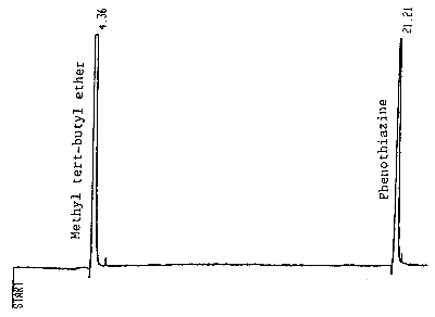
Figure 1. A standard of 165 µg/mL phenothiazine in
methyl t-butyl ether.
5. References
- 5.1. "Documentation of the Threshold Limit Values and Biological
Exposure Indices", Fifth Edition, American Conference of Governmental
Industrial Hygienists Inc., Cincinnati, OH, 1986, p. 472.
5.2. Windholz, M., "The Merck Index", Tenth Edition, Merck & Co., Rahway N.J., 1983, p. 1046.