| Method no.: | 34 |
| Matrix: | Air |
| Target concentration: | 10 ppm (18 mg/m3) (OSHA PEL) |
| Procedure: | Samples are collected by drawing known volumes of air through standard size sampling tubes containing XAD-7 resin coated with 10% NBD chloride by weight. The samples are desorbed by shaking with tetrahydrofuran (with a small amount of sodium bicarbonate present) and analyzed by high-performance liquid chromatography using a fluorescence or visible detector. |
| Recommended air volume and sampling rate: |
10 L at 0.2 L/min |
| Reliable quantitation limit: (Based on a 10-L air volume) |
24 ppb (43 µg/m3) |
| Standard error of estimate at the target concentration: (Section 4.4.) |
5.5% |
| Status of method: | Evaluated method. This method has been subjected to the established evaluation procedures of the Organic Methods Evaluation Branch. |
| Date: February 1982 | Chemist: Carl J. Elskamp |
OSHA Analytical Laboratory
Salt Lake City, Utah
1. General Discussion
- 1.1. Background
- 1.1.1. History
The current recommended sampling procedure for dimethyl amine (DMA) in the OSHA Field Operations Manual is collection in midget impingers containing sulfuric acid (Ref. 5.1.). This is also the recommended procedure for a number of other low molecular weight aliphatic amines. The analysis is normally done by gas chromatography. This sampling procedure, besides being cumbersome to use in the field has never been validated. Peak tailing, ghosting, and column decomposition due primarily to the aqueous media can make the analysis of the free amine difficult. (Ref. 5.2.) Considering the problems of the current recommended procedures, a better sampling and analysis scheme was needed. It would also be desirable that this scheme could also be used for a number of other low molecular weight amines.
In NIOSH methods (Refs. 5.3. and 5.4.) silica gel is recommended as a collection medium for aliphatic amines. Further studies by NIOSH (Ref. 5.5.) indicate that the amines are not stable on uncoated silica gel. Attempts were made by NIOSH to stabilize the amines by coating the silica gel with oxalic acid. In a recent communication with NIOSH personnel (Ref. 5.6.), it was reported that the attempt failed, presumably because the oxalic acid migrated from the silica gel. Even if a way was found to stabilize the amines, the analysis would involve direct measurement of the free amine which is difficult as previously mentioned. With these thoughts in mind, a derivative that could be formed directly on a solid sorbent sampling tube was investigated.
A number of reagents react with amines to form suitable
derivatives. These include dansyl chloride (Ref. 5.7.),
fluorescamine (Ref. 5.8.), and NBD chloride (Refs. 5.9. and 5.10.).
NBD chloride is an abbreviation for
Initial attempts to collect DMA were made by coating Gas Chrom R (firebrick, with a high surface area and few active sites) with NBD chloride and sampling a stream of DMA. This resulted in an unacceptable breakthrough volume of less than one liter for a standard size adsorbent tube. It was apparent that a sorbent must be used that would retain the amine long enough to allow it to react with the NBD chloride.
Further attempts were made by coating silica gel and Florisil. The breakthrough volumes for these coated sorbents were very high, but recoveries were very low no matter what solvent was used for desorption. Coated XAD-4 demonstrated a high capacity for DMA, but when it was coated with NBD chloride, it turned brown indicating a possible complicating reaction had taken place. XAD-7 coated with 10% NBD chloride by weight provided satisfactory breakthrough volumes and high desorption efficiencies when tetrahydrofuran (THF) was used as the desorption solvent. The tubes are stable for at least two months. It was found that when solid sodium bicarbonate was added to the desorption vial before shaking standards or samples, higher and more consistent recoveries were obtained. This is possibly due to the fact that some of the DMA may be tied up as a hydrochloride salt (hydrochloric acid is a product of the reaction between NBD chloride and DMA) and the addition of the sodium bicarbonate converts the amine salt to the free amine which can then react with the NBD chloride. It was found that by shaking the standards and samples horizontally for an hour at room temperature, enough time was allowed for a complete reaction to occur. Using coated XAD-7, a successful validation of a collection and analytical procedure for DMA was carried out. It is anticipated that other primary and secondary low molecular weight amines and ethanolamines can be collected with coated XAD-7, and analyzed in a similar manner. Future methods evaluation work will determine if a common sampling and analytical procedure for these amines is possible. Also, literature indicates (Ref. 5.11.) that some mercaptans may be done in a similar fashion, which would provide a more suitable sampling procedure and an analytical procedure that would probably be more sensitive and precise than the current method (Ref. 5.12.).
1.1.2. Toxic effects
(This section is quoted directly from the "Occupational Health Guidelines for Chemical Hazards" (Ref. 5.13.) and is for information only and should not be taken as the basis of OSHA policy.)
- "Dimethylamine gas is a severe respiratory, eye, and mucous
membrane irritant in animals. Animals repeatedly exposed to
concentrations of approximately 100 to 200 ppm for 18-20 weeks
showed marked irritation of the respiratory tract with pulmonary
edema as well as hepatic injury, including centrolobular necrosis;
corneal injury was observed in guinea pigs and rabbits after 9
days of exposure. Various spe cies survived 5 ppm of continuous
exposure for 90 days without signs of toxicity, but at autopsy
some showed mild inflammatory changes in the lungs. A drop of
undiluted dimethylamine placed on a rabbit's cornea caused the
cornea to become as white as the sclera in 1 minute. Both the
liquid and the vapor are highly irritating to the eyes and may
result in loss of visual acuity. Dermatitis and conjunctivitis are
occasionally observed in chemical workers after prolonged exposure
to the gas. No systemic effects from industrial exposure have been
reported."
1.1.3. Potential workplace exposure
Following are some common operations in which exposure to DMA may occur as reported in "Occupational Health Guidelines for Chemical Hazards." (Ref. 5.13.)
DMA is used:
- in preparation of spinning solvents for acrylic and polymeric
fibers.
as raw material in synthesis of agricultural chemicals; vulcanization accelerators for sulfur-cured rubber; softeners, lubricants; textile waterproofing agents; cationic surfactants; pharmaceuticals; detergents and soaps; as an antioxidant.
as a general solvent; acid gas adsorbent and flotation agent in manufacture of dyes and in electroplating.
as a photographic chemical, plasticizer, and ion exchange agent.
as a stabilizer in natural rubber latex.
as a stabilizer for certain types of resins (polymer ization inhibitor).
as a retarder in spinning bath of rayon (for tire cord).
as a component of rocket propellants; as an anti knock agent in other fuels.
1.1.4. Physical properties (Ref. 5.13.)
| molecular weight: | 45.1 |
| boiling point (760 mm Hg): | 6.9°C |
| specific gravity: | 0.68 (liquid at 0°C) |
| vapor pressure: | 1.72 atm at 20°C |
| color: | colorless liquid or gas |
| odor: | pungent, fish, or ammonia-like |
| flammable limits in air, % by volume: |
lower: 2.8; upper: 14.4 |
| autoignition temperature: | 402°C |
| molecular formula: | (CH3)2NH |
1.2. Limit defining parameters (The air concentrations listed throughout this method are based on an air sample volume of 10 L and a desorption volume of 2 mL.)
- 1.2.1. Detection limit of the analytical procedure
The detection limit of the analytical procedure is 2.8 ng per injection. This is the amount of DMA which will give a peak whose height is approximately 5 times baseline noise. (Section 4.1.)
1.2.2. Detection limit of the overall procedure
The detection limit of the overall procedure is 0.43 µg per sample (24 ppb or 43 µg/m3). This is the calculated amount of DMA spiked on the sampling device which allows recovery of an amount of DMA equivalent to the detection limit of the analytical procedure. (Section 4.2.)
1.2.3. Reliable quantitation limit
The reliable quantitation limit is 0.43 µg per sample (24 ppb or 43 µg/m3). This is the smallest calculated amount of analyte which can be quantitated within the requirements of at least 75% recovery and a precision (±1.96 SD) of ±25% or better. (Section 4.2.)
The reliable quantitation limit and detection limits reported in the method are based upon optimization of the instrument for the smallest possible amount of analyte. When the target concentration of an analyte is exceptionally higher than these limits, they may not be attainable at the routine operating parameters.
- 1.2.4. Sensitivity
The sensitivity of the analytical procedure over the concentration range of 5 to 20 ppm is 1090 area counts per µg DMA/mL. The sensitivity is determined by the slope of the calibration curve. (Section 4.3.) The sensitivity will vary somewhat with the particular instrument used in the analysis.
1.2.5. Recovery
The recovery of analyte from the collection medium during storage must be 75% or greater. The recovery of DMA from samples used in a 17-day storage test remained above 93% when samples were stored at ambient or refrigerated temperatures. (Section 4.4.)
1.2.6. Precision (analytical method only)
The pooled coefficient of variation obtained from replicate determinations of analytical standards at 5, 10, and 20 ppm is 0.017. (Section 4.3.)
1.2.7. Precision (overall procedure)
The overall procedure must provide results at the target
concentration that are ±25% or better at the 95% confidence level.
The precision at the 95% confidence level for a
1.3. Advantages
- 1.3.1. The solid sorbent tube provides a convenient method for
sampling.
1.3.2. DMA is analyzed as a derivative which is specific, stable, and easier to quantitate than the free amine.
1.3.3. The analysis is rapid, sensitive, and precise.
1.4. Disadvantages
- 1.4.1. The method has not been field tested.
1.4.2. Sampling tubes are not commercially available.
2. Sampling Procedure
- 2.1. Apparatus
- 2.1.1. Samples are collected by use of a personal sampling pump
that can be calibrated to within ±5% of the recommended flow rate
with the sampling tube in line.
2.1.2. Samples are collected on solid sorbent sampling tubes containing XAD-7 coated with 10% NBD chloride by weight. The tube consists of two sections of coated XAD-7 separated by a glass wool plug. The front section contains 80 mg of coated sorbent and the back section, 40 mg. The sections are held in place with glass wool plugs in a glass tube 4-mm i.d. × 70-mm length.
The coated XAD-7 is prepared by rinsing the 20/50 mesh resin several times with methyl alcohol to remove fines. The resin is extracted for 24 h with methyl alcohol in a Soxhlet Extractor and dried by vacuum. The dried resin is coated with 10% NBD chloride by weight using methylene chloride as a solvent. The solvent is removed by rotary evaporation.
2.2. Reagents
None required
2.3. Technique
- 2.3.1. Connect the sampling tube to the sampling pump with
flexible tubing. Air being sampled should not pass through any hose
or tubing before entering the sampling tube.
2.3.2. The sampling tube is placed vertically in the employee's breathing zone.
2.3.3. After sampling, the tube is sealed immediately with plastic caps.
2.3.4. Submit at least one blank for each sample set. The blank should be handled in the same manner as samples, except no air is drawn through it.
2.3.5. Record sample volume (in liters of air) for each sample, along with any potential interferences.
2.3.6. Any bulk sample(s) must be shipped in a separate container(s) from the air samples.
2.4. Breakthrough
The 5% breakthrough volume from a test atmosphere containing 20.4 ppm (37.7 mg/m3) DMA was 61.5 L, corresponding to a capacity of 2.32 mg when sampling at 0.203 L/min. (Section 4.5.)
2.5. Desorption efficiency
The desorption efficiency of DMA from spiked sample tubes is 93.6% over the range of 5 to 20 ppm. (Section 4.6.)
2.6. Recommended air volume and sampling rate
- 2.6.1. The recommended air volume is 10 L.
2.6.2. The recommended sampling rate is 0.2 L/min.
2.7. Interferences
- 2.7.1. There are no known interferences to the sampling
procedure.
2.7.2. An interference study was performed in which a 10-L sample of a test atmosphere containing 10 ppm each of DMA and methylamine was collected. There was no difference in the amount of DMA derivative found whether methylamine was present or not.
2.7.3. Suspected interferences should be reported to the laboratory with submitted samples.
2.8. Safety precautions
- 2.8.1. Attach the sampling equipment to the employee so that it
will not interfere with work performance or safety.
2.8.2. Follow all safety practices that apply to the work area being sampled.
3. Analytical Procedure
- 3.1. Apparatus (The particular apparatus used for this study can
be found in Figure 4.3.)
- 3.1.1. High-performance liquid chromatograph equipped with a
fluorescence and/or visible detector.
3.1.2. An HPLC column capable of separating the DMA derivative from NBD chloride and any interferences. A Radial CN column was used in this study in the normal phase since the NBD chloride derivatives fluoresce stronger in nonaqueous solvent systems. Reduced sensitivities with fluorescence detection will be obtained with the use of an aqueous reverse phase column system.
3.1.3. An electronic integrator or some other suitable method of measuring peak areas.
3.1.4. A mechanical shaker.
3.1.5. Volumetric flasks for preparing standards and making dilutions.
3.1.6. Pipets and syringes for preparing standards, making dilutions, and dispensing reagents.
3.1.7. Small vials with Teflon-lined caps capable of holding 3 mL.
3.2. Reagents
- 3.2.1. HPLC grade isopropanol and isooctane.
3.2.2. Reagent grade tetrahydrofuran (THF).
3.2.3. Reagent grade sodium bicarbonate.
3.2.4. Dimethylamine solution in water, of known concentration, or dimethylamine gas.
3.2.5. Reagent grade NBD chloride. (7-chloro-4-nitrobenzo-2-oxa1,3-diazole)
3.3. Standard preparation
- 3.3.1. Prepare a stock standard of DMA by diluting a known
volume of DMA with THF.
3.3.2. A working standard is prepared by injecting microliter amounts of the stock standard into 2.0 mL of a solution of NBD chloride (0.4 g NBD chloride per 100 mL THF) in a small vial. This solution will immediately turn yellow-green upon addition of the DMA.
3.3.3. Add approximately 25 mg of solid sodium bicarbonate to the vial and seal. (This is easily done by using the large end of a standard size disposable dropping pipette as a spatula.) The standards are shaken in a horizontal position for 1 h.
3.4. Sample preparation
- 3.4.1. Transfer each section of the sample to separate vials.
The glass wool plug must be added to the vials if they contain
entrapped XAD-7 beads. The glass tube is discarded.
3.4.2. Add 2.0 mL of the THF to each vial.
3.4.3. Add approximately 25 mg of sodium bicarbonate to each vial.
3.4.4. Seal the vials and shake in a horizontal position for 1 h. The vials should be positioned parallel with the shaker's movement.
3.5. Analysis
- 3.5.1. HPLC conditions
| fluorescence detector: | 465 nm excitation 525 nm emission |
| injection size: | 15 µL |
| column: | Waters Radial CN |
| solvent: | isooctane:isopropanol, 80:20 at 3 mL/ min. |
| retention time of DMA derivative: |
3.5 min |
| alternate detector: | visible at 465 nm |
| chromatogram: | Section 4.7. |
3.5.2. Peak areas are measured by an integrator or other suitable means.
3.5.3. A calibration curve is constructed from peak areas of standard injections. Sample concentrations must be bracketed by standards.
3.6. Interferences
- 3.6.1. Any compound that has the same general retention time as
the DMA derivative and responds on the detector used is an
interference. Possible interferences should be reported to the
laboratory with submitted samples by the industrial hygienist. The
derivatives of methylamine, ethylamine, and diethylamine can be
separated from the DMA derivative.
3.6.2. HPLC parameters (i.e. solvent composition, column, detector, etc.) may be changed to circumvent interferences.
3.6.3. Retention time on a single column is not considered proof of chemical identity. Samples over the PEL should be confirmed by GC/MS or other suitable means.
3.7. Calculations
The DMA concentration is obtained from the calibration curve in terms of micrograms per sample. The air concentration for samples is calculated using the following formulae. If any DMA derivative is found on the backup section, it is added to the front section. The total amount is then corrected by subtracting the total amount found in the blank.
| mg/m3 = | blank-corrected micrograms per
sample
(liters of air sampled) (desorption efficiency) |
ppm = (mg/m3)(24.46)/(45.1) = (mg/m3)(0.542)
| where | 24.46 | = | molar volume (liters) at 25°C and 760 mm Hg |
| 45.1 | = | molecular weight of DMA |
3.8. Safety precautions
- 3.8.1. Avoid skin contact and inhalation of all chemicals used,
especially DMA and NBD chloride.
3.8.2. Restrict the use of all chemicals to a fume hood if possible.
3.8.3. Wear safety glasses and lab coat at all times.
4. Backup Data
- 4.1. Detection limit of the analytical procedure
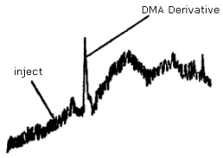
The detection limit was determined by making a 15-µL injection of a 0.187 µg/mL standard. This standard is based on the weight of DMA added to make the standard. Shown in Figure 4.1. is a chromatogram obtained from a Schoeffel FS 970 Fluorescence detector set at 0.01 µA range, 4.25 sensitivity, and 6-second time constant. The recorder was set at 0.2 cm/min and 10 mV full scale.
4.2. Detection limit of the overall procedure and reliable quantitation limit
The recovery was determined by making four 15-µL injections of extracted samples prepared by liquid injection at the analytical detection limit. The samples were allowed to set overnight before being extracted.
Detection Limit of the Overall Procedure
and Reliable Quantitation Limit Data
|
| |||
| % recovery | statistics | ||
|
| |||
| 86 86 91 81 |
SD |
= = |
86 4.1 |
|
| |||
Since samples are desorbed with 2 mL, the detection limit of the overall procedure and reliable quantitation limit is:
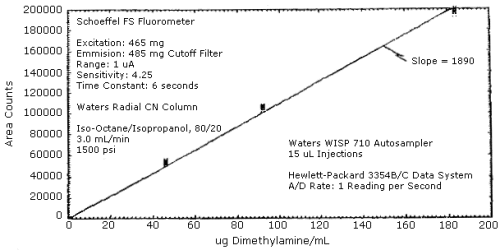
4.3. Sensitivity and precision
The sensitivity and precision of the analytical procedure were obtained from multiple injections of analytical standards. These data are given in Table 4.3. and shown graphically in Figure 4.3.
Sensitivity and Precision Data
|
| |||
| × target conc. µg/mL |
0.5× 46 |
1× 92 |
2× 184 |
|
| |||
| area
counts SD CV |
52197 53494 54319 54536 54494 55346 54064 1088.7 0.020 |
105640 106995 106494 106952 105220 102657 105660 1634.6 0.015 |
200783 197153 197846 202175 193807 196153 197986 3063.0 0.015 |
|
| |||
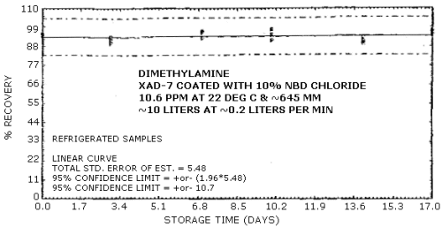
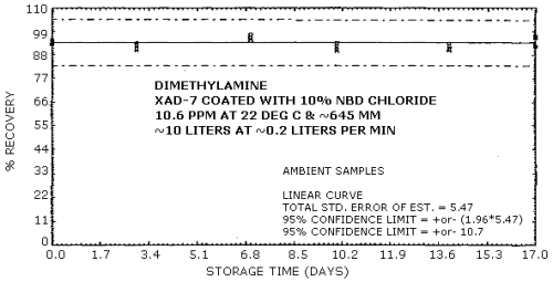
4.4. Recovery and storage data
Storage samples were generated from a test atmosphere (air) containing 10.6 ppm DMA at approximately 80% relative humidity, 22°C, and 645 mm Hg. Each sample was generated by sampling the test atmosphere at approximately 0.2 L/min for 50 min, resulting in a sample volume of about 10 L. An amount of coated XAD-7 equivalent to the front section of a standard adsorbent tube (about 80 mg) was used for each sample. After sampling, the adsorbent was transferred to separate WISP vials, capped, and stored. Six samples were extracted and analyzed immediately after generation, fifteen were stored in a closed drawer at ambient temperature, and fifteen were stored under refrigeration at 0°C.
Storage Tests
|
| |||||||
| storage time | % recovery | ||||||
| (days) | (refrigerated) | (ambient) | |||||
|
| |||||||
| 0 0 3 7 10 14 17 |
93.5 93.5 93.5 92.5 91.2 89.9 96.5 |
93.9 94.0 91.2 95.8 97.4 91.0 94.7 |
94.5 95.1 90.0 96.6 94.7 92.7 96.6 |
93.5 93.5 92.6 95.8 93.3 93.1 92.4 |
93.9 94.0 90.7 97.4 90.7 90.5 96.5 |
94.5 95.1 93.7 96.2 90.4 91.8 97.4 | |
|
| |||||||
These results are shown graphically in Figures 4.4.1. and 4.4.2.
4.5. Breakthrough
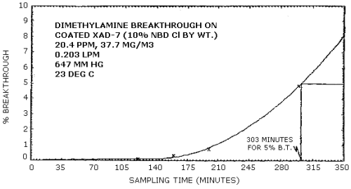
The breakthrough volume was determined from a test atmosphere containing 20.4 ppm (37.7 mg/m3) DMA. The sampling tube contained only the front section (approximately 80 mg) of adsorbent. A backup tube was connected downstream from the sampling tube. This backup tube was changed periodically and analyzed to determine the amount of DMA breaking through the sampling tube. The sampling rate used was 0.203 L/min and the test atmosphere was at 23°C, 647 mm Hg and approximately 80% relative humidity.
Breakthrough Data
|
| |||
| minutes1 | liters | mg found | % breakthrough |
|
| |||
| 60 90 120 160 200 300 |
12.2 18.3 24.4 32.5 40.6 60.9 |
0 0 0.001 0.004 0.012 0.111 |
0 0 0.11 0.33 0.78 4.84 |
|
| |||
| 1 time backup tube was changed | |||
These data are shown graphically in Figure 4.5. Breakthrough (5%) occurred at 303 min. Thus, the breakthrough volume was 61.5 L (303 min × 0.203 L/min) and the capacity was 2.32 mg (61.5 L × 2.32 mg/m3).
4.6 Desorption efficiency
The desorption efficiency was determined by injecting known amounts of a standard onto coated XAD-7 and analyzing them samples the next day. Six samples were prepared at each concentration.
Desorption Efficiency
|
| |||
| × target conc. µg/mL |
0.5× 92 |
1× 184 |
2× 368 |
|
| |||
| area
counts |
93.5 91.5 88.2 95.0 91.2 89.9 91.6 |
90.5 93.7 91.5 90.3 97.5 94.9 93.1 |
97.3 96.1 95.9 95.2 92.2 100.8 96.3 |
|
| |||
4.7. Chromatograms
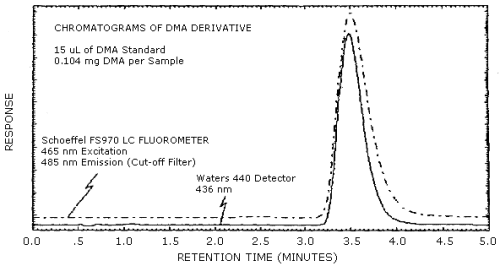
Chromatograms of a DMA standard are shown in Figure 4.7. The chromatograms are from a 15-µL injection of a 0.184 mg DMA per sample standard. The fluorescence and visible detectors were connected in series to give essentially simultaneous chromatograms. The response shown is normalized to keep the peaks about 90% full scale. This figure should not be interpreted to determine the relative response of each detector, although the detection limits for each detector are similar.
5. References
- 5.1. "Industrial Hygiene Field Operation Manual", OSHA Instruction
CPL 2-2.20, Office of Field Coordination, 1979.
5.2. Dalene, M.; Mathiasson, L.; Jonsson, J.A. J. Chromatogr (1981), 207, 37-46.
5.3. "NIOSH Manual of Analytical Methods", Vol. 1, 2nd Edition, April 1977, USDHEW, PHS, CDC, NIOSH, DHEW (NIOSH) Publication No. 77-157-A, Method P&CAM 221.
5.4. "NIOSH Manual of Analytical Methods", Vol. 3, 2nd Edition, April 1977, USDHEW, PHS, CDC, NIOSH, DHEW (NIOSH) Publication No. 77-157-C, Method S 142.
5.5. Teass, A., NIOSH (Cincinnati, Ohio), personal communication, April 17, 1981.
5.6. Teass, A., NIOSH (Cincinnati, Ohio), personal communication, October 16, 1981.
5.7. Cassidy, R. M.; LeGay, D.S.; Frei., R.W. J. Chromatog. Science (1974), 12,85.
5.8. Tomkins, B.A.; Ostrum, V.H.; Ho, C. Analytical Letters (1980), 13 (A7), 589-602.
5.9. Ghosh, P.B.; Whitehouse, M.W. Biochem J. (1968), 108, 155-6.
5.10 Klimisch, H.J.; Stadler, L. J. Chromatogr. (1974), 90, 141-8.
5.11 Nilta, K.; Bratcher, S.C.; Kronman, M.S. Biochem J. (1979), 177(2), 385-92.
5.12 Elskamp, C.J. "Methyl Mercaptan" (Method 26, Organic Methods Evaluation Branch, OSHA Analytical Laboratory, Salt Lake City, Utah). Unpublished (2-81).
5.13 "Occupational Health Guidelines for Chemical Hazards", NIOSH/OSHA, Jan 1981, DHHS (NIOSH) Publication No. 81-123.