1. General Discussion
1.1 Background
1.1.1 History of procedure
OSHA
has an exposure standard for hydroquinone at a level of 2 mg/m³ TWA.
NIOSH method 5004 collects hydroquinone on a mixed cellulose ester
filter and field extraction within one hour of collection with a 1%
acetic acid solution (Ref. 5.1). The acetic acid is to prevent
hydroquinone from isomerizing to benzoquinone. Retention studies
performed at the OSHA lab with humid air (91% RH) showed vaporization
of the hydroquinone off of the filters, with only 81% recovery on
filters analyzed immediately after the air was drawn. OSHA Method 39
recommends collection of pentachlorophenol on OVS-7 tubes and
desorption with methanol (Ref. 5.2), since hydroquinone is similar to
pentachlorophenol, this means of collection and analysis were
attempted. The hydroquinone sublimed off the glass fiber filter, and
collected on the XAD-7 resin. There it isomerized to benzoquinone in
the presence of water vapor, and the benzoquinone reacted with the
XAD-7 resin. This isomerization continued with storage, with more
benzoquinone being formed each day stored. To prevent the
isomerization of hydroquinone to benzoquinone, a phosphoric acid
coated XAD-7 resin was used for a retention study. No loss of the
hydroquinone was observed. This media was further evaluated and found
to have good retention, desorption, and storage.
1.1.2
Potential workplace exposure (Ref. 5.3)
Hydroquinone is used as
a depigmentor, a photographic reducer and developer, a reagent in
determination of small quantities of phosphates, and an antioxidant in
oils and greases. It is used as an intermediate in the manufacture of
dyes. In human medicine, it is an ingredient in topical creams for
blemishes, and in bleaching creams.
1.1.3 Toxic Effects (This
section is for information purposes and should not be taken as the
basis for OSHA policy.) (Ref. 5.4)
Hydroquinone is a skin, eye,
mucous membrane, and gastrointestinal irritant. In humans, ingestion
of 1 gram of hydroquinone has caused tinnitus, nausea, vomiting,
shortness of breath, cyanosis, convulsions, delirium, and collapse.
Death has occurred in some individuals after ingestion of 5 grams of
hydroquinone. Workplace exposure for greater than 5 years, to levels
that had no systemic effects, caused staining and opacification of the
cornea.
1.1.4 Physical properties (Ref. 5.3):
| Synonyms: |
1,4-Benzenediol; Quenelle; p-dihydroxybenzene; Hydroquinol; Aids; Black
and White Bleaching cream; Eldoquin; Eldopaque; Quinine;
Tecquinol |
| Molecular weight: |
110.11 |
| Melting point: |
170-171ºC |
| Boiling point: |
285-287ºC |
| Flash point: |
165ºC (329ºF) (closed cup) |
| Odor: |
phenolic |
| Color: |
white to light yellow crystals
which turn brown with exposure to light and air |
| Molecular formula: |
C6H6O2 |
| CAS: |
123-31-9 |
| IMIS: |
1490 |
| RTECS: |
MX3500000; 41010 |
| DOT: |
UN 2662 (Poison) |
| Structure: |
 | 1.2
Limit defining parameters
1.2.1 The detection limit of the
analytical procedure is 1 µg hydroquinone. This is the smallest amount
that could be detected under normal operating conditions.
1.2.2
The overall detection limit is 0.05 mg/m³. (All mg/m³ amounts in this
study are based on a 20 liter air volume and 1 mL desorption.)
1.3. Advantages
1.3.1 The sampling procedure is
convenient.
1.3.2 The analytical method is reproducible and
sensitive.
1.3.3 Reanalysis of samples is
possible.
1.3.4 It may be possible to analyze other compounds
at the same time.
1.3.5 Interferences may be avoided by proper
selection of column and GC parameters, or LC parameters if liquid
chromatography is used. 1.4
Disadvantages
None known.
2. Sampling procedure
2.1 Apparatus
2.1.1 A calibrated personal sampling
pump, the flow of which can be determined within ±5% at the
recommended flow.
2.1.2 XAD-7 tubes containing 20/60 mesh XAD-7
coated with 10% phosphoric acid, with an 80 mg adsorbing section and a
40 mg backup section, separated by a silane treated glass wool plug
before, after, and between the adsorbing sections. The ends are flame
sealed and the glass tube containing the adsorbent is 7 cm long, with
a 6 mm O.D. and 4 mm I.D., SKC tubes or equivalent.
2.1.3 Tubes
are available through SKC, catalog number 226-98, or can be prepared
from phosphoric acid coated XAD-7 resin, which is prepared in the
following manner.
Approximately 100 grams of Amberlite XAD-7
20/60 mesh, a porous polyacrylate adsorbent manufactured by Rolm and
Haas, was washed several, times with 100 mL deionized water to remove
the fine particles. The resin was washed three times with 100 mL
methanol, then three times with acetonitrile, and the excess
acetonitrile was removed by vacuum filtration. The resin was placed in
a round bottom flask and treated with a solution of 14 mL reagent
grade phosphoric acid in 200 mL acetonitrile. It was allowed to stand
for 10 minutes, then the resin was dried using a rotary evaporator.
The acid-coated XAD-7 resin, with the odor of acetonitrile present,
was the stored in a tightly sealed container or packed into
tubes. 2.2 Sampling
technique
2.2.1 Open the ends of the coated
XAD-7 tubes immediately before sampling.
2.2.2 Connect the
coated XAD-7 tubes to the sampling pump with flexible
tubing.
2.2.3 Place the tubes in a vertical position to
minimize channeling, with the smaller section towards the
pump.
2.2.4 Air being sampled should not pass through any hose
or tubing before entering the coated XAD-7 tubes.
2.2.5 Seal
the coated XAD-7 tubes with plastic caps immediately after sampling.
Seal each sample lengthwise with OSHA Form-21 sealing
tape.
2.2.6 With each batch of samples, submit at least one
blank tube from the same lot used for samples. This tube should be
subjected to exactly the same handling as the samples (break ends,
seal, & transport) except that no air is drawn through
it.
2.2.7 Transport the samples (and corresponding paperwork)
to the lab for analysis.
2.2.8 Bulks submitted for analysis
must be shipped in a separate mailing container from other
samples. 2.3 Desorption
efficiency
Six tubes were spiked with loadings of 4.0 µg (0.2
mg/m³), 20 µg (1 mg/m³), 40 µg (2 mg/m³), and 80 µg (4 mg/m³)
hydroquinone. They were allowed to equilibrate overnight at room
temperature. They were opened, each section placed into a separate 2 mL
vial, desorbed with 1 mL of methanol with 0.25 µL/mL dimethyl formamide
internal standard, desorbed for 30 minutes with shaking, and analyzed by
GC-FID. The overall average was 100%.(Table 2.3)
Table 2.3
Desorption
Efficiency
|
| Tube# |
4.0 µg |
% Recovered
20 µg |
40 µg |
80 µg |
|
| 1 |
100 |
101 |
103 |
103 |
| 2 |
103 |
101 |
103 |
101 |
| 3 |
102 |
99.4 |
98.0 |
98.6 |
| 4 |
102 |
97.3 |
103 |
98.1 |
| 5 |
100 |
98.7 |
98.2 |
103 |
| 6 |
100 |
100 |
95.8 |
97.6 |
|
|
|
|
|
| average |
101 |
99.6 |
100 |
100 |
| |
|
|
|
|
| overall average |
100 |
|
|
| standard deviation |
±2.19 |
|
|
| 2.4 Retention
efficiency
The six coated XAD-7 tubes were spiked with 40 µg (2.0
mg/m³) hydroquinone, allowed to equilibrate overnight, and had 20 liters
humid air (91% RH) pulled through them. They were opened, desorbed, and
analyzed by GC-FID. The retention efficiency averaged 99.7%. There was
no hydroquinone found on the backup portions of the tubes. (Table
2.4)
Table 2.4
Retention
Efficiency
|
| Tube # |
%Recovered 'A' |
% Recovered 'B' |
Total |
| 1 |
101 |
0.0 |
101 |
| 2 |
100 |
0.0 |
100 |
| 3 |
98.7 |
0.0 |
98.7 |
| 4 |
101 |
0.0 |
101 |
| 5 |
97.5 |
0.0 |
97.5 |
| 6 |
99.8 |
0.0 |
99.8 |
| |
|
|
|
| |
|
average |
99.7 |
| 2.5
Storage
Coated XAD-7 tubes were spiked with 40 µg (2.0 mg/m³)
hydroquinone and stored at room temperature, in room light, until opened
and analyzed. The recoveries averaged 97.4% for the 14 days stored.
(Table 2.5)
Table 2.5
Storage Study
|
| Day |
% Recovered |
|
| 7 |
97.6 |
| 7 |
96.6 |
| 7 |
95.7 |
| 14 |
100 |
| 14 |
98.3 |
| 14 |
96.0 |
| |
|
| overall average |
97.4 |
| 2.6
Precision
The precision was calculated using the area counts from
six injections of each standard at concentrations of 4.0, 20, 40, and 80
µg/mL hydroquinone in the desorbing solution. The pooled coefficient of
variation was 0.0388. (Table 2.6)
Table 2.6
Precision Study
|
| Injection Number |
4.0 µg/mL |
20 µg/mL |
40 µg/mL |
80 µg/mL |
| 1 |
1186 |
5715 |
11525 |
22647 |
| 2 |
1187 |
5636 |
11428 |
22860 |
| 3 |
1149 |
5650 |
11525 |
22782 |
| 4 |
1169 |
5777 |
11391 |
22745 |
| 5 |
1162 |
5750 |
11587 |
22853 |
| 6 |
1136 |
5814 |
11612 |
23155 |
| |
|
|
|
|
| Average |
1165 |
5724 |
11511 |
22840 |
| |
|
|
|
|
| Standard Deviation |
±88.4 |
70.5 |
86.8 |
173 |
| CV |
0.0759 |
0.0123 |
0.00754 |
0.00757 |
| Pooled CV |
0.0388 |
|
|
|
|
where:

A(1), A(2), A(3), A(4) = # of Injections at
each level
CV1, CV2, CV3, CV4 = Coefficients at each level
2.7 Air volume and sampling rate
studied
2.7.1 The air volume studied is 20
liters.
2.7.2 The sampling rate studied is 0.2 liters per
minute. 2.8
Interferences
Suspected interferences should be listed on sample
data sheets.
2.9 Safety precautions
2.9.1 Sampling equipment should be
placed on an employee in a manner that does not interfere with work
performance or safety.
2.9.2 Safety glasses should be worn at
all times.
2.9.3 Follow all safety practices that apply to the
workplace being sampled. 3. Analytical method
3.1 Apparatus
3.1.1 Gas chromatograph equipped with
a flame ionization detector. A HP 5890 gas chromatograph was used in
this study.
3.1.2 GC column capable of separating the analyte
and an internal standard from any interferences. The column used in
this study was a 15-meter DB-WAX capillary column, 0.25 µm df, and
0.32 mm I.D.
3.1.3 An electronic integrator or some other
suitable method of measuring peak areas.
3.1.4 Two milliliter
vials with Teflon-lined caps.
3.1.5 A 10 µL syringe or other
convenient size for sample injection.
3.1.6 Pipettes for
dispensing the desorbing solution. The Glenco 1-mL dispenser was used
in this method.
3.1.7 Volumetric flasks - 5 mL and other
convenient sizes for preparing standards.
3.1.8 An analytical
balance capable of weighing to the nearest 0.01 mg.
3.1.9 If
liquid chromatography is used for analysis the instrumentation is a
liquid chromatograph equipped with an autosampler and an ultraviolet
detector, and a C18 column. A Waters 510 pump, 710 B WISP, 440
detector with extended wavelength module were used. The column was a
Supelco LC-18-DB, 5µ, 25 cm × 6 mm. 3.2 Reagents
3.2.1 Purified GC grade nitrogen,
hydrogen, and air. (for GC analysis only)
3.2.2 Hydroquinone,
Reagent grade
3.2.3 Methanol, HPLC grade
3.2.4 Dimethyl
formamide, Reagent grade
3.2.5 Desorbing solution is 0.25 µL/mL
dimethyl formamide internal standard in methanol. If analysis is
performed by liquid chromatography, it may be better to not use an
internal standard, as on many C18 columns it is difficult to separate
the dimethyl formamide from the hydroquinone.
3.2.6 Deionized
water (for mobile phase for LC analysis only)
3.2.7 Phosphoric
acid (for mobile phase for LC analysis only) 3.3. Sample preparation
3.3.1 Sample tubes are opened and each
section of each tube are placed in separate 2 mL vials, along with the
separating glass wool.
3.3.2 Each section is desorbed with 1 mL
of the desorbing solution.
3.3.3 The vials are sealed
immediately and allowed to desorb for 30 minutes on a shaker, a
roto-rack, or a sample rocker. 3.4 Standard preparation
3.4.1 Standards are prepared by
diluting a known quantity of hydroquinone with the desorbing
solution. Stock solutions also had 0.1 mL/L of
H3PO4 added to them.
3.4.2 At least two
separate stock standards should be made. Dilutions of the stock
standards are prepared to bracket the samples. For this study,
standards ranged from 1 to 80 µg/mL. 3.5 Analysis
3.5.1 Gas chromatograph
conditions.
| Flow rates (mL/min) |
Temperature (ºC) |
| Nitrogen (makeup): 30 |
Injector: 240 |
| Hydrogen (carrier): 1.5 |
Detector: 240 |
| Air: 450 |
Column: 80º for 2 min |
| Hydrogen (detector): 30 |
then 10ºC/min to 200ºC |
| Injection size: 1 µL |
|
| Elution time: 15.69 min |
|
| Chromatogram: |
|

Figure 1. An analytical standard of 40 µg/mL
hydroquinone in methanol with 0.25 µL/mL dimethyl formamide internal
standard, and analyzed by gas chromatography.
3.5.2. Liquid
chromatograph conditions.
| Column: |
5 µ Supelco LC-18-DB, 6
× 250 mm |
| Mobile
Phase: |
1 mL of
0.1:5:95 phosphoric acid:methanol:water (if using methanol with
the dimethyl formamide internal standard) |
| Mobile
Phase: |
1 mL/min of 0.1:25:75
phosphoric acid:methanol:water (if using methanol only as
desorbing solvent) |
| Injection size: |
10 µL |
| Detector: |
UV at 219 nm (UV max is
223 nm, secondary max is 200 nm, tertiary max is 291 nm) (note:
if the analysis is performed at 219 the DMF can be used as an
internal standard, but if 291 nm is used the DMF does not have a
response at that wavelength) |
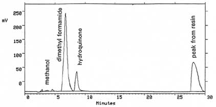
Chromatogram:

Figure 2. An analytical standard of 40 µg/mL
hydroquinone in methanol with 0.25 µL/mL dimethyl formamide internal
standard, analyzed by liquid chromatography with an UV detector at 219
nm, and using a mobile phase of 0.1:5:95
H3PO4:methanol:water.

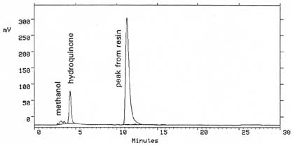
Figure 3. An
analytical standard of 40 µg/mL hydroquinone in methanol with 0.25
µL/mL dimethyl formamide internal standard, analyzed by liquid
chromatography with an UV detector at 219 nm, and using a mobile phase
of 0.1:25:75 H3PO4:methanol:water.
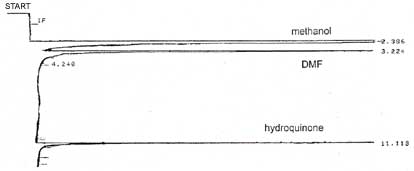
The
following chromatogram was analyzed on an HP5890 Series II gas
chromatogram with a flame ionization detector. The column was a
60-m 0.32-mm i.d. capillary column with a 0.25 µm DB-1 film
thickness. The temperature program was 80°C for 4 min the
10°C/min to 160°C hold 5 min, with the injector at 200°C and the
detector at 250°C.

Figure 4. A chromatogram of 75 µg/mL hydroquinone in
methanol with DMF internal standard.
3.5.3 Peak areas are
measured by an integrator or other suitable means.
3.6 Interferences
(analytical)
3.6.. Any compound having the general
retention time of the analyte or the internal standard used is an
interference. Possible interferences should be listed on the sample
data sheet. GC parameters should be adjusted if necessary so these
interferences will pose no problems.
3.6.2 Retention time data
on a dingle column is not considered proof of chemical identity.
Samples over the target concentration should be confirmed by GC/Mass
Spec or other suitable means.
3.6.3 There is a reaction between
the excess phosphoric acid and the methanol to form trimethyl
phosphate. On the column used in this study trimethyl phosphate eluted
at 10 minutes. The amount formedis approximately 50 µg. If another
column is used for this analysis, the trimethyl phosphate should be
separated from the other peaks. 3.7 Calculations
3.7.1 A curve with area counts versus
concentration is calculated from the calibration
standards.
3.7.2 The area counts for the samples are plotted
with the calibration curve to obtain the concentration of hydroquinone
in solution.
3.7.3 To calculate the concentration of analyte in
the air sample the following formulas are used:
(µg/mL)(desorption volume)
desorption efficiency |
= |
mass of analyte in
sample |
mass of analyte in sample
air volume in liters |
= |
µg/L of analyte |
| mg³ of analyte |
= |
µg x mg x 1000 L
L x 1000 µg x m³ |
3.7.4 The above equations can be consolidated to
form the following formula. To calculate the mg/m³ of analyte in the
sample based on a 20 liter air sample:
| mg/m³ |
= |
(µg/mL)(DV)(mg)(1000 L)
(20 L)(DE)(1000 µg)(m³) |
where:
| µg/mL |
= |
concentration of analyte in sample
or standard |
| DV |
= |
Desorption volume |
| 20 L |
= |
20 liter air sample |
| DE |
= |
Desorption
efficiency |
3.7.5 This
calculation is done for each section of the sampling tube and the
results added together. 3.8
Safety precautions
3.8.1 All handling of solvents should
be done in a hood.
3.8.2 Avoid skin contact with all
chemicals.
3.8.3 Wear safety glasses, gloves and a lab coat at
all times. 4.
Recommendations for further study
Collection study should be
performed. 5. References
5.1 "NIOSH Manual of Analytical
Methods", U.S. Department of Health and Human Services, Public Health
Service, Centers for Disease Control, National Institute for
Occupational Safety and Health, Third Edition, Method 5004.
5.2
Cummins, K., Method 39, "Phenol and Cresol", Organic Method's Evaluation
Branch, OSHA Salt Lake Technical Center, 1982.
5.3 Windholz, M.,
"The Merck Index", Eleventh Edition, Merck Co., Rahway N.J., 1989, p.
762.
5.4 "Documentation of the Threshold Limit Values and
Biological Exposure Indices", Fifth Edition, American Conference of
Governmental Industrial Hygienists Inc., Cincinnati, OH, 1986, p.
319. |

